Lumbar disc herniation

Example of a L4-5 lumbar disc herniation on MRI (magnetic resonance imaging).
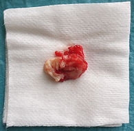
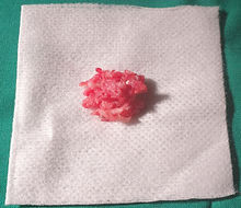
This is what surgical disc herniation looks like. After the removal of such hernia, the spinal nerve roots are completely released, thus the pain disappears.

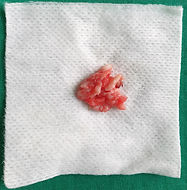
A very big disk hernia
evacuated 3 x 1.5 cm
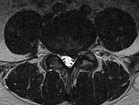
Synovial cyst


A L4-5 level synovial cyst is visible before the operation


There is no synovial cyst visible after the operation
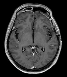
Meningioma
Meningioma at the frontal cranial base before surgery


There is no meningioma after the operation
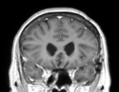
Falx cerebri meningioma
Falx cerebri meningioma between the hemisheres


There is no meningioma after surgery
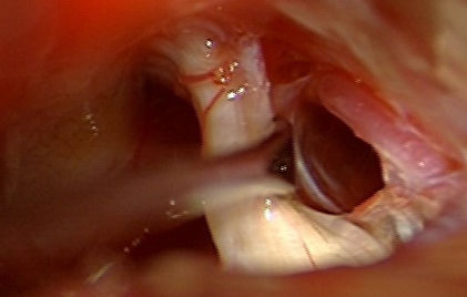
Trigeminal neuralgia
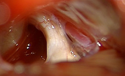
The neurovascular conflict between the trigeminal nerve and an artery is visible - the nerve is being irritated by the artery in arachoid adhesions. Thus the shooting facial pain is generated.
_JPG.jpg)
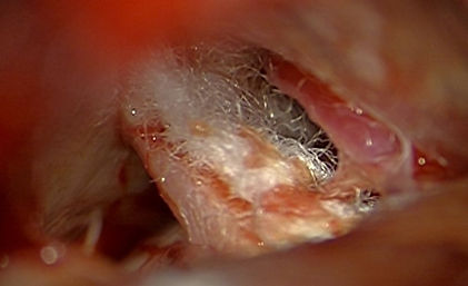
The adhesions are cut and the vessel is moved away from the nerve.
_JPG.jpg)
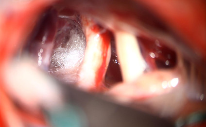
An additional teflon sponge as a cushion is positioned between the nerve and artery. The artery no more irritates the nerve and the facial pain disappears.
Trijzaru nerva neiralģija 2. gadījums

Redzams neirovaskulārs konflikts - artērijas cilpa
iespiežas nervā (a. cerebelli superior)
Artērija tiek aizvirzīta prom un starpā tiek ievietota telfona starplika. Sāpes sejā izzūd.
Trijzaru nerva neiralģija 3. gadījums


Spinal tumour
_JPG.jpg)
The tumour is located in front of the spinal cord
and pushes the cord backwards
_JPG.jpg)
The tumour is being removed

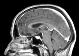
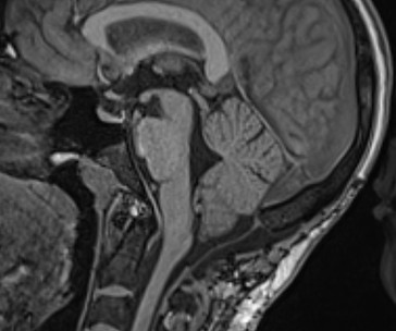
Chiari I malformation

Preoperativa image. Lowering of the cerebellar tonsils and compression of the brainstem is visible. The cerebrospinal fluid flow is compromised.
Postoperative image. Free space around cerebellar tonsils is visible now, no more compression. Free flow of the cerebrospinal fluid has been obtained.
Glioblastoma - malignant brain tumour
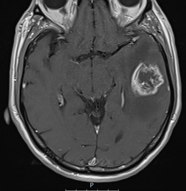
Malignant brain tumour visible on the preoperative image (arrow).

The postoperative cavity is visible after the removal of the tumour (arrow).

5-ALA (Gliolan) fluorescence use during the operation. The tumor cells glow red in the blue light (arrow).
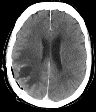
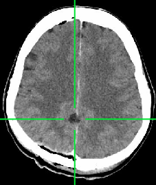
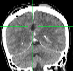
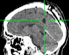
Spontaneous intracerebral haemorrhage (haematoma)

Intracerebral haemorrhage is visible.

Postoperative scan- the blood clot has been removed
ETV - endoscopic 3rd ventriculostomy

An opening has been made at the bottom of the 3rd ventricle in a case of occlusive hydrocephalus -
the cerebrospinal fluid is freely pulsating.
Hydrocephalus has been cured.
Cerebral metastasis

Cerebral MTS is visible before the operation